Source: SQA / Qualifications Scotland. This is a StemInteract HTML companion, not an official HTML edition. Use the original images for figures, graphs and table layout.
PDF page 1
Selectable page text
AH
National
Qualications
2026
X807/77/11 Biology
Supplementary sheet
TUESDAY, 28 APRIL
9:00 AM – 12:00 NOON
Supplementary sheet for question 1
*X8077711*
A/PB
© Qualifications Scotland 2026PDF page 2
Selectable page text
1. Marine mammals display several physiological adaptations to their marine environment.
   Their cells contain the molecule myoglobin, which can associate with oxygen in a similar
   way to haemoglobin, providing an oxygen store within cells. Higher myoglobin
   concentrations in the muscles of marine mammals allows them to increase their internal
   oxygen stores, increasing the time available to dive.

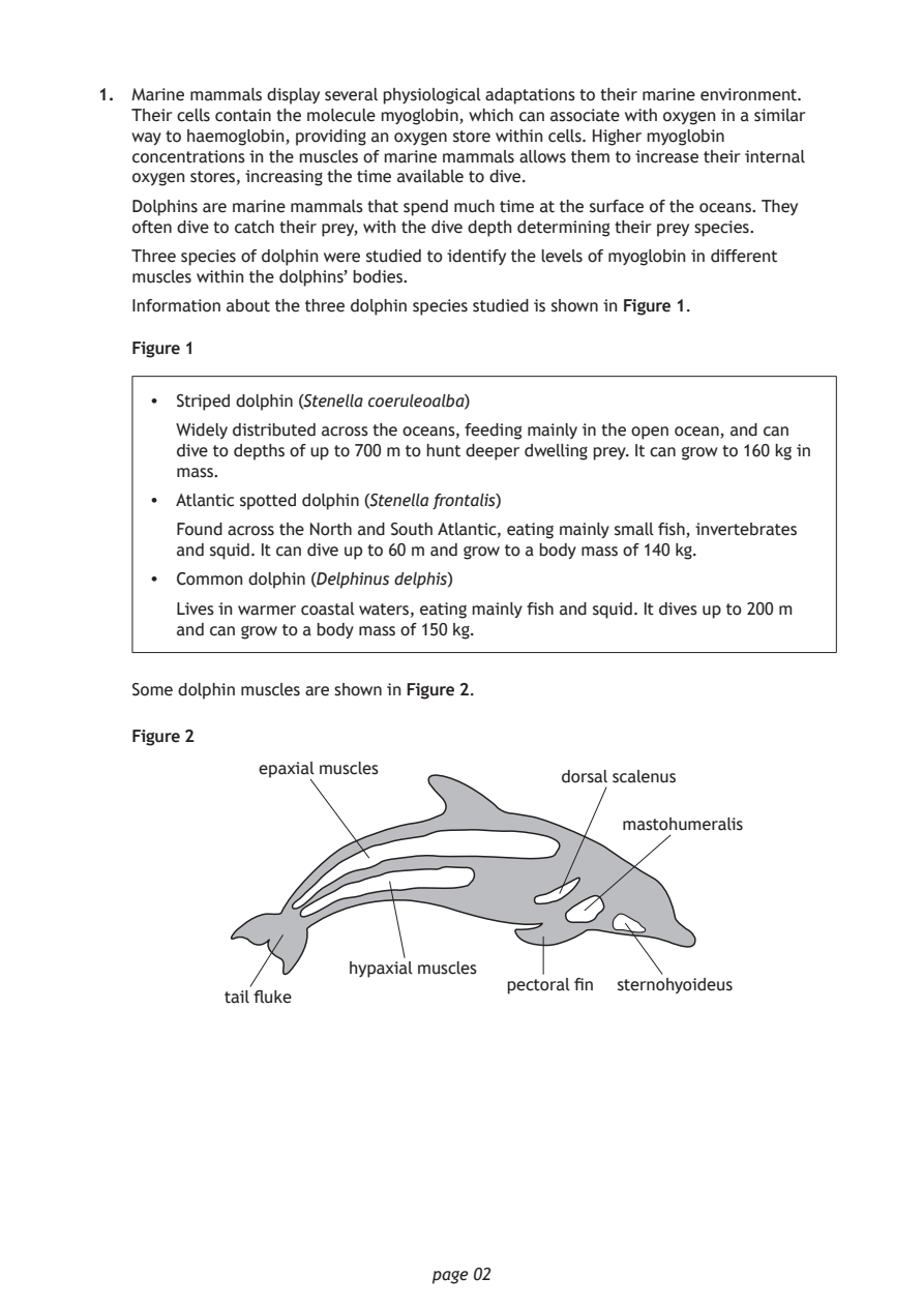
   Dolphins are marine mammals that spend much time at the surface of the oceans. They
   often dive to catch their prey, with the dive depth determining their prey species.
   Three species of dolphin were studied to identify the levels of myoglobin in different
   muscles within the dolphins’ bodies.

   Information about the three dolphin species studied is shown in Figure 1.

   Figure 1

    •  Striped dolphin (Stenella coeruleoalba)
       Widely distributed across the oceans, feeding mainly in the open ocean, and can
       dive to depths of up to 700 m to hunt deeper dwelling prey. It can grow to 160 kg in
       mass.
    •  Atlantic spotted dolphin (Stenella frontalis)

       Found across the North and South Atlantic, eating mainly small fish, invertebrates
       and squid. It can dive up to 60 m and grow to a body mass of 140 kg.
    •  Common dolphin (Delphinus delphis)

       Lives in warmer coastal waters, eating mainly fish and squid. It dives up to 200 m
       and can grow to a body mass of 150 kg.

   Some dolphin muscles are shown in Figure 2.

   Figure 2

              epaxial muscles
                                         dorsal scalenus
                                              mastohumeralis

                      hypaxial muscles
                                    pectoral fin sternohyoideus
           tail fluke

                             page 02
PDF page 3
Selectable page text
1. (continued)

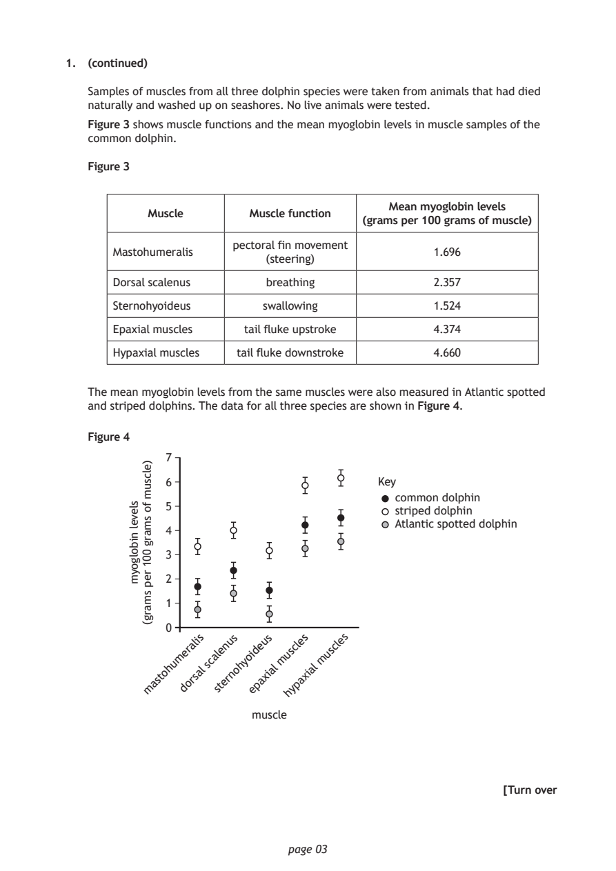
   Samples of muscles from all three dolphin species were taken from animals that had died
   naturally and washed up on seashores. No live animals were tested.

   Figure 3 shows muscle functions and the mean myoglobin levels in muscle samples of the
   common dolphin.

   Figure 3

                                           Mean myoglobin levels
           Muscle       Muscle function
                                       (grams per 100 grams of muscle)
                      pectoral fin movement
      Mastohumeralis                             1.696
                          (steering)
      Dorsal scalenus     breathing              2.357
      Sternohyoideus      swallowing             1.524

      Epaxial muscles   tail fluke upstroke      4.374

      Hypaxial muscles tail fluke downstroke     4.660

   The mean myoglobin levels from the same muscles were also measured in Atlantic spotted
   and striped dolphins. The data for all three species are shown in Figure 4.

   Figure 4

             7
             6

                                            common dolphin
             5
                                            striped dolphin
                                            Atlantic spotted dolphin
             4
             3
             2

             1
             0

                             page 03
        slevel
        nibolgoym
          )elcsum
          fo
          smarg
          001
          rep
          smarg(
                                         Key
                 i s u s u s  e s   e s
                    n    e
          m a s ^{t} o h u m d e o r ^{a} r s ^{l} a ₗ s c a s l t e e r n o h y o e ⁱ d p a x ⁱ a ₗ m u s y ^{c} p ^{l} a x ⁱ ₐₗ m u s ^{c} ^{l}
                             h
                         muscle
                                                          [Turn over
PDF page 4
Selectable page text
1. (continued)

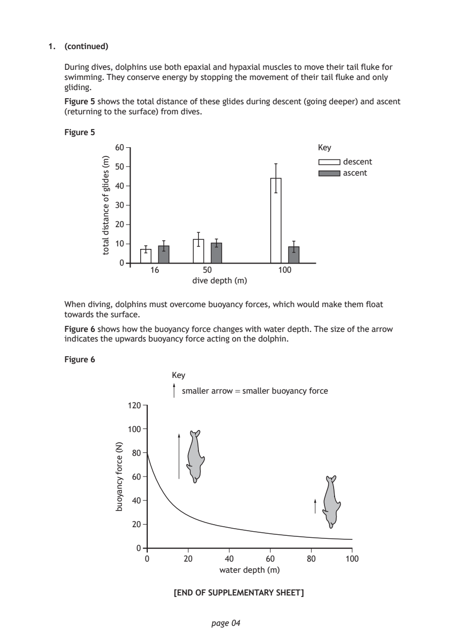
   During dives, dolphins use both epaxial and hypaxial muscles to move their tail fluke for
   swimming. They conserve energy by stopping the movement of their tail fluke and only
   gliding.

   Figure 5 shows the total distance of these glides during descent (going deeper) and ascent
   (returning to the surface) from dives.

   Figure 5

            60
                                                     descent
            50
                                                     ascent
            40
            30

            20

            10

             0

                          dive depth (m)

                             page 04
          )m(
          sedilg
          fo
          ecnatsid
          latot
                                                 Key
                  16        50            100
   When diving, dolphins must overcome buoyancy forces, which would make them float
   towards the surface.
   Figure 6 shows how the buoyancy force changes with water depth. The size of the arrow
   indicates the upwards buoyancy force acting on the dolphin.
   Figure 6
                      Key
                        smaller arrow = smaller buoyancy force
              120
              100
               80
               60
               40
               20
                0
                 0      20      40     60      80     100
                               water depth (m)
            )N(
            ecrof
            ycnayoub
                       [END OF SUPPLEMENTARY SHEET]